Lesson 1 of the Six Lessons Approach to Biomimetic Dentistry relates to Diagnosis and Treatment of Caries. The first lesson primarily revolves around the concepts of the Peripheral Seal Zone (PSZ) and Caries Removal Endpoints (CRE). Both of these terms were first coined in the 2012 paper by Alleman and Magne.
Removal of Caries from the Peripheral Seal Zone (PSZ)
The Peripheral Seal Zone (PSZ) is the 2-3 mm of tooth structure (enamel, DEJ, and superficial dentin) in which complete caries and crack removal is indicated.
Caries Detecting Dye is used to ensure that no caries is present 1-2 mm inside of the DEJ.
Bond strengths of 30-50 MPa can be achieved in the PSZ.
Master Biomimetic Dentistry Online.
Increase predictability. Reduce stress.
Davey Alleman, DMD
Removal of Deep Caries Using Caries Removal Endpoints (CRE)
After achieving complete caries removal in the Peripheral Seal Zone (PSZ), Caries Removal Endpoints (CRE) are used to partially remove deep caries over the pulp while avoiding pulp exposure.
Caries Removal Endpoints are measured using a periodontal probe along the long axis of the tooth:
- Horizontally – 3 mm measured interproximally from the adjacent tooth’s marginal ridge.
- Vertically – 5 mm measured occlusally from the cavo-margin.
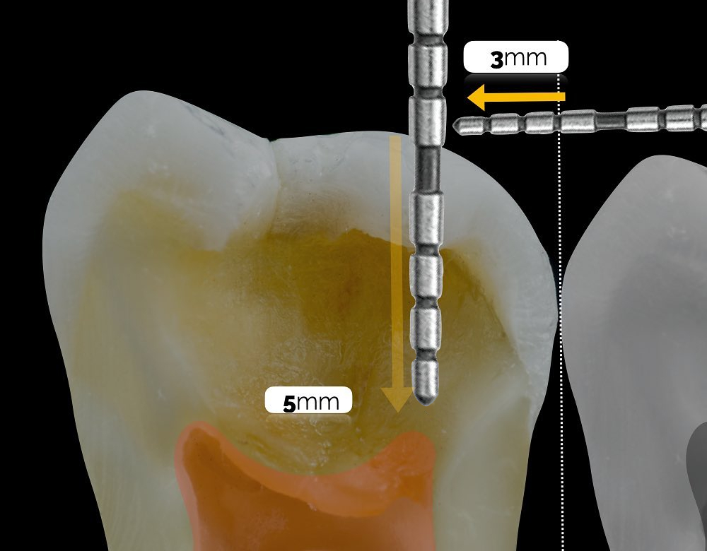
Photo Credit: Dr Andres Celi
Caries removal is stopped within these boundaries, even if the Caries Detector Dye (CDD) is staining red or pink (further information regarding CDD can be found below). The area over the pulp, encircled by the PSZ, in which partial caries removal is indicated, is known as the Central Stop Zone (CSZ).
Assuming a caries-free PSZ has been created, the high resulting bond strength in the PSZ allows the deep caries over the pulp to be sealed, therefore avoiding pulp exposure and allowing a Biomimetic restoration to be created.
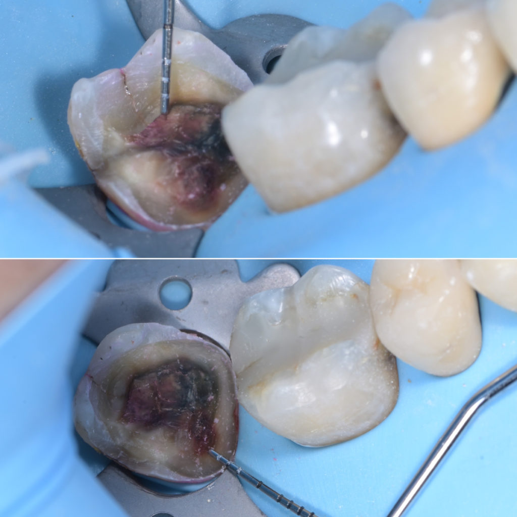
Photo Credit: Dr John Weselake
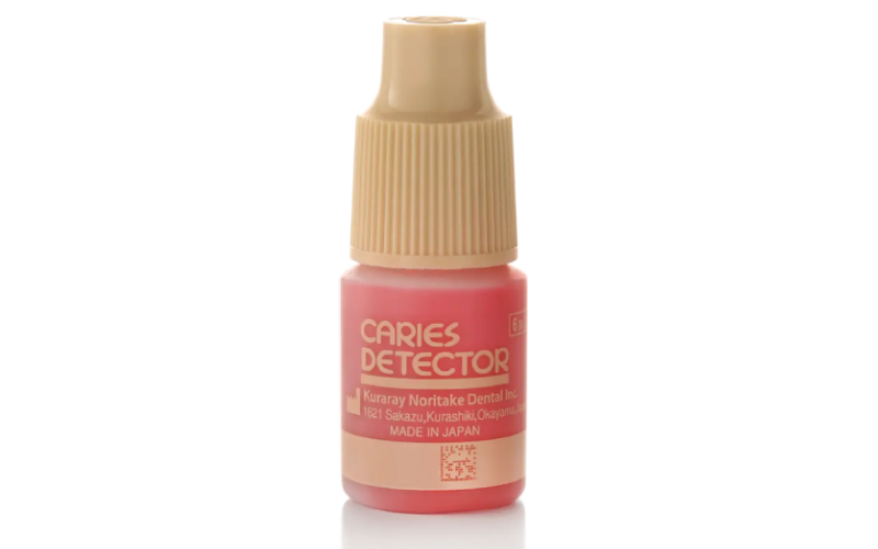
Caries Detector Dye
Using tactile or visual methods to assess for caries is subjective. If using an explorer to check if the surface of dentin is ‘hard’ to probe and therefore free from caries – how hard is ‘hard’? These techniques can be unreliable.
A more objective method of assessing for caries is by using Caries Detector Dye (CDD). CDD works by staining denatured collagen a certain colour – usually red or green depending on the product used. It commonly consists of 1% Acid Red solution in a propylene glycol solvent. The collagen fibers of outer carious dentin are loosened, and therefore allow penetration of the solvent and staining of the substrate.
Outer carious dentin (irreversibly denatured infected dentin) stains darker (red), whereas inner carious dentin (reversibly denatured affected dentin) stains lighter (pink). This is important as different substrates have different bonding potentials. This is commonly known as the Hierarchy of Bondability (HOB).
How to Use Caries Detector Dye
- Gain initial access to the carious lesion as appropriate
- Clean the carious lesion using water and then air dry thoroughly
- Apply one drop of CDD using a microbrush and allow to penetrate for 10 seconds
- Rinse with water and then air dry
- Remove caries following the PSZ and CRE concepts
- Repeat as necessary until CRE achieved.
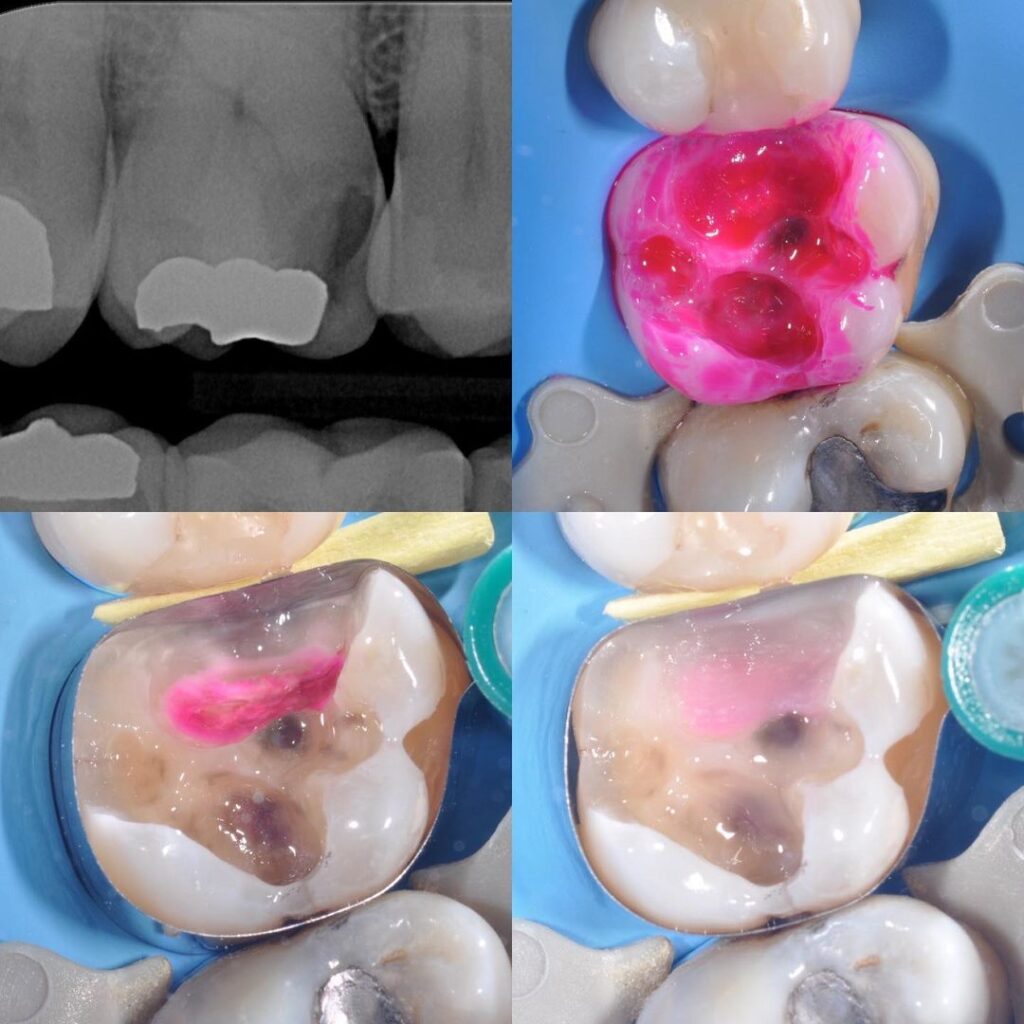
Photo Credit: Dr Jorge Diaz
Ready to take your Biomimetic Dentistry to the next level?
Click here to access our online, on-demand Biomimetic Dentistry courses. Stay at the forefront of modern Dentistry by learning from international leaders.
Relevant Literature
- Alleman DS, Magne P. A systematic approach to deep caries removal end points: the peripheral seal concept in adhesive dentistry. Quintessence Int. 2012 Mar;43(3):197-208.
- Anderson MH, Charbeneau GT. A comparison of digital and optical criteria for detecting carious dentin. J Prosthet Dent. 1985 May;53(5):643-6.
- Fusayama T. Clinical guide for removing caries using a caries-detecting solution. Quintessence Int. 1988 Jun;19(6):397-401.
- Yoshiyama M, Tay FR, Doi J, Nishitani Y, Yamada T, Itou K, Carvalho RM, Nakajima M, Pashley DH. Bonding of self-etch and total-etch adhesives to carious dentin. J Dent Res. 2002 Aug;81(8):556-60.