A key term of biomimetic restorative dentistry, clearly defined. Part of The Hybrid Layer's free reference glossary.
Glossary term
Free to read
Summary – What is a Peripheral Rim Fracture (PRF)?
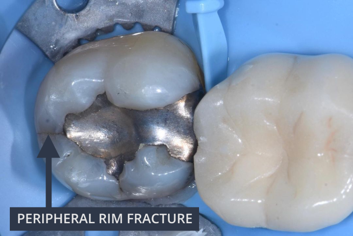
If a traditional cavity is prepared with an isthmus width >2 mm, the tooth is no longer connected in a biomimetic way. The tooth can flex in a range of around 200 microns which results in stress concentration. Fracture resistance decreases and the marginal ridges are more likely to crack, leading to Peripheral Rim Fractures (PRF).
Fig 1. Clinical example of a Peripheral Rim Fracture (PRF). Photo credit: Dr Pete Butkus
Learn More…
To learn more about Peripheral Rim Fractures in relation to structural compromises and micromovements, see Lesson 2 of the Six Lessons Approach to Biomimetic Dentistry below.
Three lectures on the fractures behind structural compromise: biomimetic cracked-teeth diagnosis and treatment, crack dynamics in dental hard tissues, and crack treatment options and prevalence.
Learn biomimetic dentistry from world-leading experts.
Watch the Peripheral Rim Fracture and every other biomimetic concept demonstrated on real cases by the clinicians who use them every day. One membership, every lecture, worldwide.
55+ hours of on-demand lectures from 35+ international experts
Verifiable CPD/CE certificates with every completed course
The Biomimetic Learning Pathway so you always know what to watch next
Free, practical biomimetic dentistry tips, straight to your inbox. Sign up and you will get some of our most popular tips each day over the next week, then a new tip every Thursday.
You're in. Welcome.
Check your inbox. You'll receive one of our most popular tips each day over the next week, then a new biomimetic tip every Thursday.
The Hybrid Layer
Welcome back
Sign in to continue your learning and pick up your CPD where you left off.